|
|
| |
|
What is fluorescence?
The CytoScience SMR fluorescence microscope offers a highly sophisticated high-powered microscope for disease diagnosis and general fluorescence microscopy, made from the best materials at a most competitive price. No sacrifice has been made to the quality but the costs have been lowered through design and application of new technologies. Microscopes have previously been thought of as sensitive instruments. Due to the simplicity in design and extreme ruggedness, it is now possible to carry out fluorescence microscopy not only in general laboratories, but also in general practice, which results in considerable time and cost saving.
Until recently these microscopes have been expensive and difficult to use. Now, fluorescence microscopy is the choice for high contrast imaging and diagnostics. In well equipped laboratories fluorescence has become the gold standard.
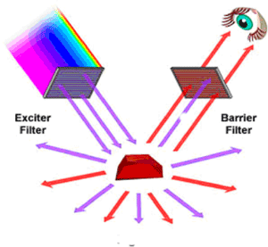
With specific labelling (dyes, antibodies) it is possible to image down to the level of specific molecules. The principle is quite simple. Excitation light is passed through a filter to produce monochromatic light of a short wavelength. This excites the dye in the object, which fluoresces. The light emitted is separated from the scattered light by a barrier filter.
The SMR uses the most common and most efficient method, called epi-fluorescence, to get the excitation light onto the object by deflecting the excitation light onto the specimen through the objective and using this as the condenser. This is done by means of a dichromatic mirror.
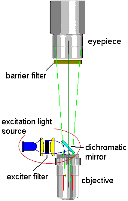
The use of the dichromatic mirror allows the excitation light to be focussed into the objective while allowing the emmision ligfht to pass through. This method gives the best contrast to the image. A second filter – the barrier further reduces scattered excitation light to further imprive contrast. We have designed the specification filters to give the best results in terms of excitation power and to pass as much emmision light as possible. The barrier filter is briad band so that a wide spectrum of light – from green to red is passed through. This combination allows the use of a very large nunber iof commion dyes. 90 % of all applications can be applied using these optics.
One of the many innovations in the SMR is the use of a LED. CytoScience is the first microscope manufacturer to introduce LED lighting for fluorescence microscopy. Conventional fluorescence microscopes use expensive, power consuming, short life span, non regulatable and very fragile gas discharge lamps (such as Xenon- or Mercury-lamps). A blue LED was first used for low magnification (x10). CytoScience conducted further development on this LED technology to provide much higher power and better focussed light to enable high magnification (up to x60) fluorescence microscopy. The use of the LED as a light source enables CytoScience to lower the cost of the microscope and at the same time improve the instrument’s power consumption, easily regulatable light intensity and especially size and robustness.
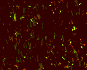
An example of the achievable high contrast is shown below. It shows a population of bacteria (E.coli) that has been treated with a sterilising agent. The process was not complete so some of the bacteria are still living (green) and some are dead (red). Only with fluorescence, one can distinguish between live and dead cells. (The live strain is labelled with Syto Green and the dead stain with Propridium Iodide)
|
|
|
