|
|
| |
|
VIRUSES
Some viruses can be diagnosed using fluorescence microscopes. Usually microscopes cannot resolve the structure or detect easily the presence of virus particles in blood. Where the tissue contains enough virus particles a labelled antibiody can be used to detect the virus in the tissue. This is the case for Rabies for example, Below we show an example of Dengue virus labelled with a specific FITC coupled antibody.
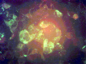
This photomicrograph was taken on a Cytoscience SMR microsocpe at the Bundeswehr Iinstitute of Microbiology.
Information on Dengue can be obtained at http://www.cdc.gov/ncidod/dvbid/dengue/
being a virus, a microscope cannot resolve the stucture or detect easily the presence in blood.
By selectively attacking one of the cells of the immune system – mainly the CD4+ T cell and macrophages through their receptors, the virus enters a race betweent he bodies ability to produce these cells and the viruses rate of killing them. The infected person can take years to die, until the body is exhausted, and die due to reduced immune responses to other organisms, such as TB. The diagnosis done by detecting antibodies to the virus but the surveillance of the disease progress is not done by measuring the viral load but by an absolute count of the CD4+ cells per volume of blood.
Enumeration technologies recommended by the WHO are found at the following link http://www.who.int/diagnostics_laboratory/publications/en/cd4_is_draft.pdf
Since the CSD4+ count needs fresh blood, the only strategy that can work effectively is to do this at the site of the point of care. A rising CD4+ count indicates the treatment is working effectively. The cost of monitoring can be more expensive than the antiviral drugs. |
|
|
